
Muscles of the Head, Trunk and Arms


(Page numbers refer to Pictorial Anatomy of the Cat, rvsd, by Gilbert.)
We will be studying head and trunk muscles of the cat, most of which are analogous to those in the human. Working with a skinned cat ( see previous protocol ) remove cutaneous muscle layer (allows cat to twitch its skin) and a white layer of superficial fascia to better see muscle fiber directions and make the muscles more apparent. Carefully outline, separate and lift the muscles by use of a blunt probe. If the structure in question has multiple fibers in it, it is muscle. Look for intersections between fiber directions, this often indicates two muscles. Fingers are the best blunt probes…
When you need to cut separated superficial muscles to see deep muscles, the superficial muscle to be reflected should be snipped midway between insertion and origin, and laid back to its origin and insertion, noting where they are located. Make four illustrations:
1) ventral thorax, upper appendage and abdomen, superficial
2) Ventral thorax and upper appendage, deep
3) Dorsal (back) deep and dorsal superficial
4) Second illustration of the deep dorsal muscles.
VENTRAL NECK, CHEST AND ABDOMEN:
(See Gilbert, p. 18)

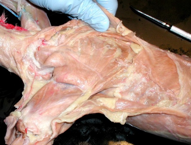
Undissected chest. Remove as much adipose tissue and fascia as you can so that the fibers of the muscles can be seen.
Can you find the pectoantebracialis, pectoralis major, latissimus dorsi and triceps brachii?
Here is the same image with the chest muscles labeled.


Lift deltoid and pectoantebrachialis as a unit and cut and reflect.


Here is a labeled view of the ventral surface of the upper appendage.
Separate pectoralis major from pectoralis minor, cut both, reflect to see: (Gilbert p. 24)
Here is a labeled version of the deep muscles of the chest and scapula.


Identify the external muscles of the abdomen (p. 24)


BACK: (p. 22)
Caution: the trapezius is very thin and easily torn when outlining it with the probe. Remove cutaneous muscle layer, note the boundary between trapezoid and the latissimus dorsi which plunges below it.
1. Lift trapezius from underlying latissimus dorsi.


2. Cut and reflect trapezius to see muscles related to or on the scapula: (p. 25). Here is a labeled view of the deep muscles of the back and scapula.


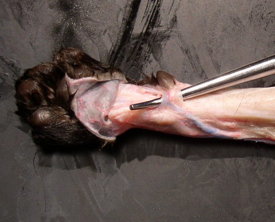
splenius capitis (to the left and below the tip of the probe)
Seen below the rhomboideus muscles.
The “bandage” muscle in the posterior neck.
Origin: upper thoracic spinous processes.
Insertion: mastoid process. process
Here again is a labeled view of the deep muscles of the back and scapula.
Muscles of the shoulders and arms
Brachial plexus: To be studied Winter Quarter:


deltoid and cutting the latissimus dorsi so that it can be reflected:


Cutting the pectoralis. The trapezius has been cut and reflected to show the scapula and rhomboideuses


