
Histology of a Leaf Cross Section


This lab is designed to be used as a means of reviewing the use of the binocular microscope.
Slide: Sun Leaf Pear, B 669a
Scan the entire leaf section using the 4x objective , note major visible features, especially vascular tissue bundles and leaf tissue structure. Find a well-defined vascular bundle (not the central vein), then rotate the 10x objective into place. Finally, examine the vascular bundle with the 40x objective. Note the various classes of cells which you can distinguish. Illustrate the 400x view containing the following labeled features which should be familiar to you from first year biology:
epidermis: epidermal cells, cuticle, stoma, guard cells
mesophyll: palisade parenchyma, spongy parenchyma, intercellular space
vascular tissues: vascular bundle, bundle sheath, xylem (large, few) and phloem (small and numerous

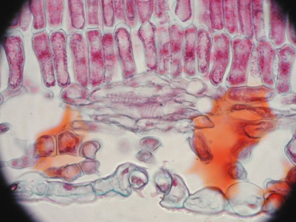
Cross section of pear leaf at the central vein, 40x

Cross section of pear leaf central vein, 100x

Vascular bundle, showing xylem (larger red cells) and phloem (indistinct small greenish cells) wrapped in bundle sheath.

Smaller vascular bundle with pallisade parenchyma and spongy parenchyma clearly shown.
Here is a labeled version of the image of the histology of the leaf.

guard cells guarding stoma at center bottom.

guard cells guarding stoma at center bottom.
Here is a labeled version of guard cells and tissues.
guard cells guarding stoma at center bottom
