
Hematocrit Protocol


SAFETY NOTE: To reduce the risk of transmission of blood-borne diseases such as AIDS and hepatitis B, wear protective gloves when handling other peoples blood, dispose of all blood contaminated materials in the provided containers, and clean up thoroughly when finished.
The number of erythrocytes in the blood must be high enough to carry sufficient O2 to the peripheral tissues, and yet not so great as to adversely increase the blood’s viscosity. A simple test to determine the percent of formed cells in blood, 99% of which are RBCs, is the hematocrit (Hct) (hemato- = blood, -crit = separate). A fresh sample of blood is introduced into a capillary tube (coated with heparin to prevent clotting), the end sealed with a putty, and the tube centrifuged to sediment the cells. The straw colored supernatant is the plasma, the RBCs sink to the bottom, and the WBCs are seen as a thin buffy coat at the top of the RBC column. By determining the percent of the total represented by the packed cells, the percent of RBCs in whole blood can be determined.
Normal Hct values for men are 40 to 54 percent, those for females, 37 to 47 percent. Anemia is defined as Hct counts below these. Anemia may be due to inadequate nutrition, blood loss, hemolytic disease, or exposure to agents which inhibit mitosis (such as radiation or chemotherapeutic agents).
[This protocol may performed simultaneously with white and red blood cell counts and blood typing.]
APPARATUS AND MATERIALS REQUIRED:
70% EtOH
Cotton balls, or Kimwipes
Sterile lancets (or Autolet apparatus with sterile fittings)
Hematocrit tubes, heparinized
sealing putty (Crit-o-seal)
Clinical centrifuge fitted with hematocrit head
Micro-capillary reader

1. Set up all apparatus required for the variety of blood tests you wish to perform (i.e., blood typing slide setup, and for blood counts: dilution pipettes, diluents, hemocytometer.)
2. Wash hands well with soap and water.
3. Swab a less-used finger (i.e. ring finger on non-writing hand) with 70% EtOH. Lance finger tip with quick firm jab to the side of the fingertip pad, wipe off first drop with clean dry cotton ball. Dispose of the used lancet safely in sharps container.
4. Fill capillary tube to within 1-2 cm of top with blood by slightly tipping it to allow blood to flow down-hill into tube. Avoid bubbles.
5. Holding tube horizontally, press filled end into sealing putty (Crit-o-seal) to plug end. It can then be stored vertically in the Crit-o-seal tray until ready to be centrifuged. (Continue collecting the rest of the samples require for your exercises before clotting occurs.)

6. Lie the tube in the centrifuge Hct head with plugged end to the outside, note the number of your slot. Ensure that a balancing hematocrit tube is placed opposite, either by someone placing their tube there, or by adding an empty tube


7. Securely screw down top of head. Turn on centrifuge to speed 6, run for 5 minutes, turn off.


8. When rotation has stopped, remove tube, note appearance.
9. Place in hematocrit reader, determine % of blood as formed cells, according to instructions on the reader. Enter your data into class data table. Illustrate the centrifuged hematocrit tube in your book and label: hematocrit tube, putty plug, packed cells (indicate % of total blood volume) and plasma.

10. Place centrifuged tube with putty towards center.

11. Align upper portion of putty with the black line.

12.Rotate assembly so that the pin stops, lining up with the 100 percent mark.
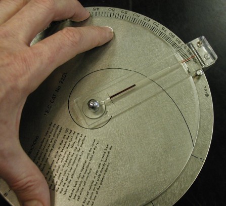
13. Rotate the upper disk to move the curved line with the top of the plasma.

14. Here the curved is lined up with the top of the plasma.

15. Rotate the entire assembly until the curved line is lined up with the boundary between the packed cells and the plasma. Read the percent packed cells off the disc at the right. (It reads 44.9 %)
