Milk Fermenters

Milk is extremely perishable and many means have been developed to preserve it. The earliest one which has been used for many thousands of years is fermentation. Milk can be fermented by inoculating fresh milk with the appropriate bacteria and keeping it at a temperature which favors bacterial growth. As the bacteria grow, they convert milk sugar (lactose) to lactic acid. You can detect its presence by the tart or sour taste (sour is how we taste acid). The lowered pH caused by lactic acid preserves the milk by preventing the growth of putrefactive and/or pathogenic bacteria which do not grow well in acid conditions.

FERMENTATION

Fermentation is a means by which cells growing anaerobically can still generate a little ATP. Fermentation is defined biochemically as the catabolism of glucose (or other sugars) in which the terminal hydrogen acceptor is an organic molecule (carbon containing). During the breakdown of sugar, known as glycolysis, excess hydrogen atoms are generated and must be deposited somewhere. In lactic acid bacteria, they “dump” excess hydrogens on to pyruvic acid, the end product of glucose. This turns pyruvic acid into lactic acid.

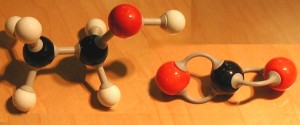
Our muscles do the same thing, which causes the sting in over exercised muscles. In all fermentation, NADH gives up its hydrogen to produce NAD, which is required for further glycolysis. Yeast too performs fermentation, but with different terminal hydrogen acceptors (acetaldehyde) and products (CO2 and ethanol). You will note that alcoholic fermentation is also an anaerobic process. Since the terminal hydrogen acceptor in each of these microbiological processes is an organic molecule, they are, by definition, fermentation.

In contrast, respiration uses an inorganic terminal hydrogen acceptor (such as oxygen). If oxygen is the acceptor, then water is produced.

Casein, the predominant protein in milk, is soluble at a neutral pH, but insoluble in acid. Thus when milk sours, casein precipitates which thickens the product. Numerous strains of bacteria are capable of converting lactose to lactic acid. We will look at several fermented milk products to study their morphology and staining characteristics.

  1. Make a thin smear of each milk product well spaced on the same slide, labeling with a wax pencil Y, B and S. (see protocol Smear and Staining of Bacterial Specimens)
  2. Stain them according to the procedure for the Gram stain (see related protocol Gram Stain Protocol), or any simple stain such as methylene blue, should you only be interested in seeing bacterial morphology.
  3. View the stained smear at 400x to determine the characteristic features, select a field which is well spread and typically stained. Then switch to 1000x with oil. (The oil immersion lens is challenging to novices. Do not use this lens unless you have been instructed in its use.)
  4. Illustrate typical fields for each milk product showing all observed morphologies of bacteria. Label the morphologies and their probable identities according to the following type of bacteria expected in these fermented milk products:

YOGURT

Yogurt is produced by a mixed culture of two types of bacteria. Imbedded in particles of the protein casein, you will see chains of cocci or diplococci (Streptococcus thermophilus) and big rod-shaped bacilli (either Lactobacillus acidophilus or L. bulgaricus). If you do a Gram stain, the bacteria will be Gram positive (purple) and the protein will be pink. The illustrations at the top of the page are micrographs I took of a Gram stain of yogurt. The purple rods are Lactobacillus, the purple spheres are Streptococcus. The pink globs are casein, milk protein.
BUTTERMILK

Buttermilk is the fermentation of milk by a culture lactic acid-producing Streptococcus lactis plus Leuconostoc citrovorum which converts lactic acid to aldehydes and ketones which gives it its flavor and aroma.

SOUR CREAM

Sour cream is produced by the same bacteria as buttermilk, but the starting milk product is pasteurized light cream. Bacteria are less numerous than in buttermilk.

PROKARYOTIC ANATOMY

TFC, 7th p 77-99, Alcamo, p. 87-, Atlas, pp 111-139, TFC, 8th p 76-96, Black 6th: 77-89, Bauman 2nd, 55-65

CELL SHAPE: coccus, bacillus, spirillum, pleomorphic

ARRANGEMENT: depends on how cells stick together after mitosis: diplo, strepto-, tetrad, sarcinae, staphylo-

GLYCOCALYX (sugar + cup, flower) sticky, gelatinous layer, shown by negative stain. (Not in spirillum)
capsule if firmly attached to bacterial cell (see p 61)
slime layer so called if loose, unorganized
Extracellular Polymeric Substance = EPS, if composed of sugars: used for attachment especially
mucopolysaccharide:   polysaccharide [dextran] cross-linked with small peptides

Function: buffers, protects fr phagocytosis, dehydration, adheres organism to substrate
pathogenicity: B. anthracis + S. pneumoniae only pathogenic if encapsulated (smooth)
adherence:       Klebsiella sticks to respiratory tract, S. mutans in forming dental caries.

“Ropy” milk, beer due to Alcaligenes viscolactis capsular material, ropy bread to B subtilis

Quellung reaction (swelling): swelling of capsule from specific antibody attack, used to type strains

FLAGELLA: (p 62) element of motility, spins to move. Flagella cannot be seen unless coated with dye.
filament is a polymer of flagellin (H antigen), hook at proximal end, attached via basal body to bacterium:
Gm pos: single pair of rings in basal body
Gm neg: two pairs of rings in basal body, inner ring rotates, outer is stator
Example:
Styles of flagellar arrangement (p63)
peritrichous                  “around hairs”                                         E. coli, etc
monotrichous               (single polar flagellum)                           Vibrio
lophotrichous               [tuft hair] (2 flagellae or more at l end)   Pseudomonas
amphitrichous              (tufts of flagellae at both ends)                Spirillum
permit chemotaxis        attractant: steady runs, repellant: runs with many tumbles.

AXIAL FILAMENTS: (p 63) in spirochetes, cell wraps around axial filament move by boring or snake-like movement (Treponema & Leptospira)

MOVEMENTS: (p 64)
Run: 
clockwise rotation, 1 second (favorable conditions)
Tumble: counterclockwise, 0.1 sec. (Unfavorable conds.)

PILI: [hair] (p 65) hollow structures of subunits pilin [antigenic]
1) fimbriae      aid in attachment to substrate, enhance pathogenicity, eg: Neisseria gonorrhoeae pellicle: fuzzy ort shiny layer on top of air-water interface by aerobic bacteria
2) sex pili aid in transfer of DNA

PROKARYOTIC ANATOMY II: CELL WALLS, PLASMA MEMBRANE, ENDOSPORES

TFC, 7th p 77-99, Alcamo, p. 87-, Atlas, pp 111-139, TFC, 8th p 76-96, Black 6th: 90-95, Bauman 2nd,65-92

CELL WALL: (p 66)
20x norm pressure inside (300 psi!), cell wall hold contents in, gives structure & protection
composed of peptidoglycan (murein), a polysaccharide with alternating NAG-NAM (10 to 65 in row):
glycan: NAG:   N acetylglucoseamine (p 66) NAM:  N-acetylmuramic acid, tetra peptide cross linking anchors cross connected via 1-5 AA (alternating D and L) with peptide cross-linking side chains.bacteria grouped according to antigenicity of cell wall (example: group A strept.)

Overview: p 68:
Gram positive     25 nm thick of peptidoglycan plus antigenic teichoic acid (p 68) (PO4 + glycerol (or ribitol) or phospholipid)
Gram negative    only 3 nm thick wall, plus second membrane on exterior of cell wall with lipopolysaccharide periplasmic space between membranes: protective enzymes

LIPOPOLYSACCHARIDE (LPS, in outer membrane of Gram negative bacteria) consists of:

Lipid A (p. 68)        embedded in outer membrane, released as endotoxin on death. Trigger macrophages to release cytokines: chills, fever, weakness, generalized aches, shock, death.
O polysaccharide   projects out, constitutes “O” antigen as in O157:H7 of pathogenic E. coli. p 68: contrast Gm +/Gm-:           As a result of the extra barrier in Gm- bacteria, more resistant to antibiotics, salts and dyes

AGENTS AGAINST CELL WALLS:

         Lysozyme               (found in tears, etc) cuts polysaccharide
         Penicillin                inhibits synthesis, leads to protoplast formation in growing cells

Mycoplasma have no cell wall, instead have cholesterol in membrane (a unique group of bacteria)

PLASMA MEMBRANE: (p 69) 60% protein, 40% phospholipid, fluid mosaic model
Mesosomes: invaginations of plasm membrane, form septum. DNA attached here incr absorb Alcohols, quaternary ammonium compounds and polymyxins damage the plasma membrane


MOVEMENT ACROSS MEMBRANE: (p 71)
Diffusion, osmosis (p 73), hypertonic solutions draw water out, cell collapses
Facilitated diffusion, active transport (p 74)

CYTOPLASM: Inclusions (stored nutrients), classification of which aid in classification:

volutin: stored poly P04 (red with methylene blue, diagnostic of C.orynebacterium diphtheria) metachromatic with methylene blue staining (variable staining)
polysaccharide starch or glycogen, iodine shows as black/purple grains
lipid:poly β-hydroxybutyric acid, sudan dyes shows (p 75)

ENDOSPORES: (p 547)    specialized resting cells, survive extreme heat , toxins, chemicals, xeroduric. Survives 10,000 yr, possibly 25 mil in amber? Four genera, only Bacillus and Clostridium are pathogenic

Illustrate process of sporulation:(p. 76)

  1. spore septum forms by invagination of plasma membrane
  2. newlysynth DNA surrounded (forespore) by double layer of membrane (spore and cell memb)
  3. Cortex of peptidoglycan between two membranes
  4. sporecoat of protein synthesized on outside.

Layers: 1) spore coat, 2) outer memb, 3) cortex, 4) inner memb, 5) inside: dipicolinic acid, stabilizes DNA


EUCARYOTIC CELLS [Not covered?]

Flagella and cilia

Cell wall

Plasma membrane

Cytoplasm

ORGANELLES: Nucleus, endoplasmic reticulum, golgi complex, mitochondria,

Centrosome and centrioles, chloroplasts, inclusions